Davson-Danielli Sandwich Model (1930)
•Proposed layers of protein adjacent to the phospholipid bilayer
•Evidence
–Proteins appear dark in electron micrographs and phospholipids appear light
•Evidence
–Proteins appear dark in electron micrographs and phospholipids appear light
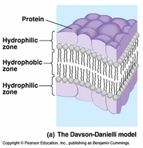
http://avonapbio.pbworks.com/w/page/9429341/Davson-Danielli%20Model
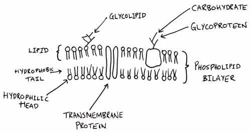
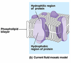
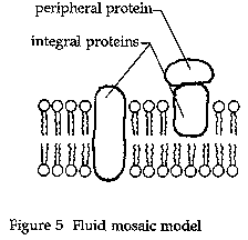
Singer-Nicolson Fluid Mosaic Model
It retains the basic lipid bilayer structure first proposed by Gorter and Grendel and modified by Danielli and Davson and Robertson. The proteins, however, are thought to be globular and to float within the lipid bilayer rather than form the layers of the sandwich-type model.
The hydrophobic tails of the phospholipids, the major lipid component of the membrane, face inward, away from the water. The hydrophilic heads of the phospholipids are on the outside where they interact with water molecules in the fluid environment of the cell. Floating within this bilayer are the proteins, some of which span the entire bilayer and may contain channels, or pores, to allow passage of molecules through the membrane. The entire membrane is fluid
•Evidence
–1. Freeze etched images of the center of membranes was interpreted as trans membrane proteins
– 2. Improvements in biochemical techniques led to discovery of new membrane structures. Membranes were found to be varied in size and shape.
|
|
|
source: http://www1.umn.edu/ships/9-2/membrane.htm
Key points
- Particles move across membranes by simple diffusion, facilitated diffusion, osmosis and active transport.
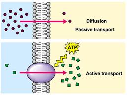
Passive transport
Doesn't use chemical energy (ATP)
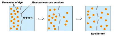
1. simple diffusion
Passive movement of particles from a region of high concentration to a region of low concentration (down a concentration gradient)
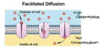
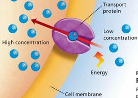
2. facilitated diffusion
Passive movement of particles from a region of high concentration to a region of low concentration (down a concentration gradient), through a selectively permeable membrane, facilitated by carrier proteins. Carrier proteins are integral proteins that allow some molecules to pass through
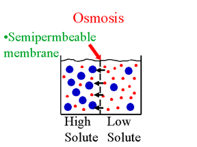
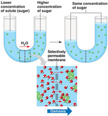
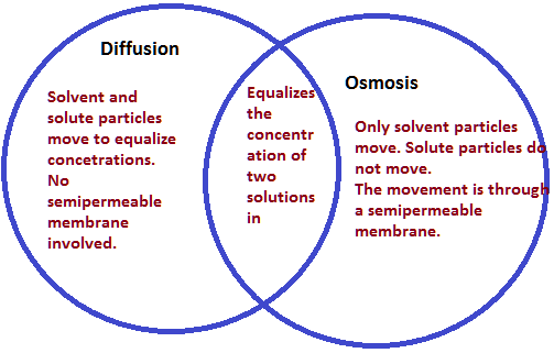
3. osmosis
note: Solutions consist of solutes dissolved in a solvent. Concentration is the measure of the amount of solute in the solution
Is the passive movement of water molecules from a region of low solute concentration (high water concentration) to high solute concentration (low water concentration) through a selectively permeable membrane
Is the passive movement of water molecules from a region of low solute concentration (high water concentration) to high solute concentration (low water concentration) through a selectively permeable membrane
|
|
compare diffusion and osmosis
active transport
Uses chemical energy (ATP) to move molecules against a concentration gradient, using membrane protein pumps. It moves the concentrations from a region of lower to a region of higher concentration.
|
|
- The fluidity of membranes allows materials to be taken into cells by endocytosis or released by exocytosis. Vesicles move materials within cells.
endocytosis and exocytosis
-Exocytosis: is the export of macromolecules from the cell. It has 4 stages:
1. The protein of the ribosomes enters the rough ER
2. Protein then exits the ER and enters the cis side (receiving side) of the Golgi apparatus
3. In the Golgi apparatus, the protein is modified and exits through the trans side (discharging side) inside the vesicle
4. The vesicle with the modified protein moves to and fuses with the plasma membrane goes out of the cell.
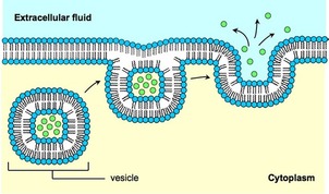
-Endocytosis: is when macromolecules enter the cell. It occurs when a portion of the plasma membrane pinches off to enclose the macromolecules. This causes a change in the shape of the membrane; a vesicle is formed and then enters the cytoplasm of the cells. After this process the membrane goes back to its original shape due to its fluid nature.
Some terms we should know:
Phagocytosis: is the ingestion of solid molecules
Pinocytosis: is the ingestion of liquid and solutes
Vesicle transport: exocytosis of protein via Golgi apparatus
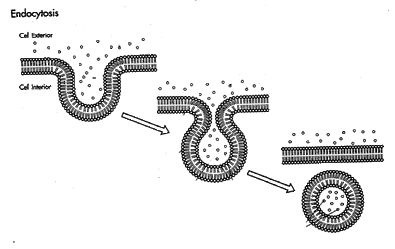
Membrane Fluidity: unsaturated hybrocarbon tail of phospholipids have kinks that keep the molecules from packing together, enhancing membrane fluidity. Cholesterol reduces membrane fluidity by reducing phospholipid movement at moderate temperatures but it also hinders solidification at low temperatures.
Liposomes for drug delivery: artificially produced vesicles that can be used for transport of drugs around the body. Good method of targeting cancer
source: Biology class
Phagocytosis: is the ingestion of solid molecules
Pinocytosis: is the ingestion of liquid and solutes
Vesicle transport: exocytosis of protein via Golgi apparatus
Membrane Fluidity: unsaturated hybrocarbon tail of phospholipids have kinks that keep the molecules from packing together, enhancing membrane fluidity. Cholesterol reduces membrane fluidity by reducing phospholipid movement at moderate temperatures but it also hinders solidification at low temperatures.
Liposomes for drug delivery: artificially produced vesicles that can be used for transport of drugs around the body. Good method of targeting cancer
source: Biology class
| 3._membranes_1.pptx |
